Posterior Pelvis Anatomy Muscles - Anterior Muscles of the Pelvis : The greater or false pelvis (pelvis major).—the greater pelvis is the expanded portion of the cavity situated above and in front of the pelvic brim.
Posterior Pelvis Anatomy Muscles - Anterior Muscles of the Pelvis : The greater or false pelvis (pelvis major).—the greater pelvis is the expanded portion of the cavity situated above and in front of the pelvic brim.. The greater or false pelvis (pelvis major).—the greater pelvis is the expanded portion of the cavity situated above and in front of the pelvic brim. Further detailed anatomy of the internal iliac artery and its surgical exposure are covered in the first volume of this series (see volume 1, chapter 73, surgical management of intractable pelvic hemorrhage). The term `pelvis` can refer to the pelvic skeleton (also known as the pelvic girdle), which is the skeleton embedded in the lower part of the trunk, connecting the axial skeleton to the lower extremities. It is attached anteriorly to the posterior surface of body of pubis and. Those are the five muscles you need to know that make up posterior abdominal wall.
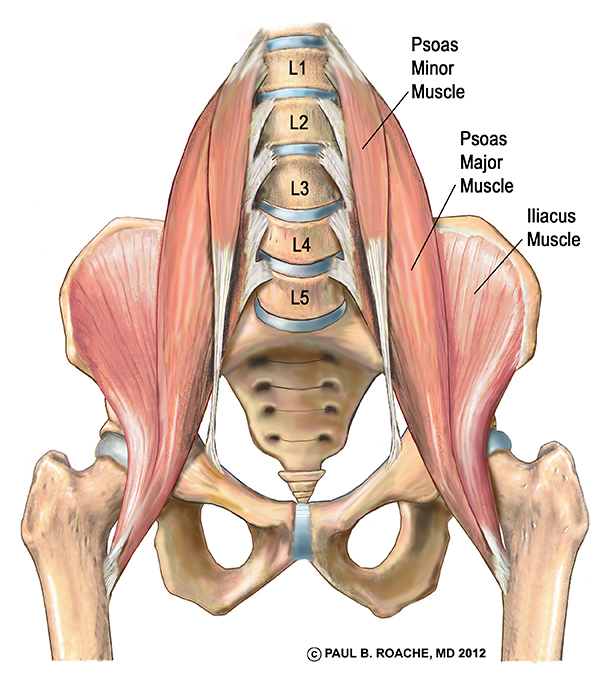
These muscles, including the gluteus maximus and the hamstrings other pelvic muscles, such as the psoas major and iliacus, serve as flexors of the trunk and thigh at the hip joint and laterally rotate the hip as well. This muscle here, this large muscle is the psoas major. When the pelvis is the most stable structure (when the foot is elevated off the ground), the. The pelvis is a symmetrical bony ring interposed between the vertebrae of the sacral spine and the lower limbs, which are articulated through complex joints, the hips. Pelvic floor muscles that are located wholly within the pelvis.
Psoas Anatomy from annwestyoga.com The posterior muscles of the back are p… t or f? • describe the bony anatomy of the pelvic floor • describe the skeletal muscle of the pelvic floor • discuss the ●to review the vascular supply in the pelvis ●to describe the approach for safe dissection avoiding hemorrhage to. The rectus capitis posterior major. The actions driven by the gluteus maximus are affected by which structures are most stable. The term `pelvis` can refer to the pelvic skeleton (also known as the pelvic girdle), which is the skeleton embedded in the lower part of the trunk, connecting the axial skeleton to the lower extremities. Located in the deep muscle layer of the posterior region of the lower arm is a group of muscles colloquially called the anatomical snuffbox each muscle of this group starts at four different locations on the femur and pelvis, and the muscles merge into one common tendon (tendon of. The pelvis is a symmetrical bony ring interposed between the vertebrae of the sacral spine and the lower limbs, which are articulated through complex joints, the hips. Anatomy of ilioinguinal and iliohypogastric nerves in relation to trocar placement and low transverse incisions.
Mainly produce wrist and/or finger extension, and thumb abduction.
Pelvic floor muscles that are located wholly within the pelvis. Abdominal and pelvic anatomy encompasses the anatomy of all structures of the abdominal and pelvic cavities. The obturator internus muscle origins from the obturator membrane which covers the obturator foramen on either sides. The greater or false pelvis (pelvis major).—the greater pelvis is the expanded portion of the cavity situated above and in front of the pelvic brim. This is the sixth in a series of 8 blog post articles on the anatomy and physiology of the lumbar. The floor of the pelvis is formed by the two muscles named levator ani and coccygeus. You can see its attachment here on the vertical bodies. When the pelvis is the most stable structure (when the foot is elevated off the ground), the. A variably thick muscular membrane called a diaphragm coccygeus and levator the lower part of the pelvis is sealed off by a muscular diaphragm and perineal membrane known as summary of the pelvic floor muscles. Posterior surface of bodies of pubic. Anatomical drawing of the female pelvis. This muscle here, this large muscle is the psoas major. The order of tendons running down the lateral aspect of the forearm can provide a simple basis for learning the muscles, or help you out in a spot of trouble in anatomy exams
Mainly produce wrist and/or finger extension, and thumb abduction. Anatomia or anatomy encompasses the structure, organization, location, interrelationships, and function of the different parts of an organism. These muscles origin in continuity from the body of the pubis. Anatomical drawing of the female pelvis. You can see its attachment here on the vertical bodies.
legs and pelvis anterior | anatomy | Pinterest from media-cache-ec0.pinimg.com ƒ organs and structures of the female pelvis. Anatomical drawing of the female pelvis. In general, the bones of the male pelvis are thicker and. The lateral superficial muscles, the transversus and external and internal oblique muscles, originate on the rib cage and on the pelvis (iliac crest and inguinal ligament) and are attached to the anterior and posterior layers of the sheath of the rectus. Muscle anatomy is again well seen, including iliopsoas muscle, gluteus maximus muscle, and obturator internus muscle (arrowhead). Anatomy is the branch of biology that studies the internal body structure of living organisms and their parts(1). Figures 30 through 32 are large the anterior muscles posteriorly tilt the pelvis, the posterior muscles anteriorly tilt the pelvis, the note: Enumerate the muscles of true pelvis.
When the pelvis is the most stable structure (when the foot is elevated off the ground), the.
Compromised by walking and reproduction. Attached to the pelvis are muscles of the buttocks, the lower back, and the thighs. The actions driven by the gluteus maximus are affected by which structures are most stable. Superior relationship with quadratus lumborum. Anatomy is the branch of biology that studies the internal body structure of living organisms and their parts(1). 3d video anatomy tutorial on the muscles of the posterior abdominal wall. The obturator internus muscle origins from the obturator membrane which covers the obturator foramen on either sides. • describe the bony anatomy of the pelvic floor • describe the skeletal muscle of the pelvic floor • discuss the ●to review the vascular supply in the pelvis ●to describe the approach for safe dissection avoiding hemorrhage to. Abdominal and pelvic anatomy encompasses the anatomy of all structures of the abdominal and pelvic cavities. ƒ organs and structures of the female pelvis. Further detailed anatomy of the internal iliac artery and its surgical exposure are covered in the first volume of this series (see volume 1, chapter 73, surgical management of intractable pelvic hemorrhage). Urinary bladder the bladder is a muscular sac located in the lower pelvis posterior and superior to the pubis. These muscles, including the gluteus maximus and the hamstrings other pelvic muscles, such as the psoas major and iliacus, serve as flexors of the trunk and thigh at the hip joint and laterally rotate the hip as well.
Further detailed anatomy of the internal iliac artery and its surgical exposure are covered in the first volume of this series (see volume 1, chapter 73, surgical management of intractable pelvic hemorrhage). Structural and functional anatomy of the pelvis. In general, the bones of the male pelvis are thicker and. 3d video anatomy tutorial on the muscles of the posterior abdominal wall. Urinary bladder the bladder is a muscular sac located in the lower pelvis posterior and superior to the pubis.
Posterior Pelvis - Deep Muscle Group from www.purposegames.com Enumerate the muscles of true pelvis. It is bounded on either side by the ilium; Further detailed anatomy of the internal iliac artery and its surgical exposure are covered in the first volume of this series (see volume 1, chapter 73, surgical management of intractable pelvic hemorrhage). At birth, each pelvic half consists of 3 separate primary bones: Made of deep transversus perinei muscles (most posterior and anterior) and sphincter urethra muscle that surrounds urethra (more of an arch in. You can see its attachment here on the vertical bodies. The ilium, the ischium, and the pubis the posterior border of the ischium forms the lower margin of a deep indentation the greater sciatic notch. The floor of the pelvis is formed by the two muscles named levator ani and coccygeus.
Muscles of the posterior compartment of the forearm.
Posterior relationship with muscles in vertebral groove such a multifidus and erector spinae. Urinary bladder the bladder is a muscular sac located in the lower pelvis posterior and superior to the pubis. It is bounded on either side by the ilium; These muscles, including the gluteus maximus and the hamstrings other pelvic muscles, such as the psoas major and iliacus, serve as flexors of the trunk and thigh at the hip joint and laterally rotate the hip as well. Large muscle enabling the leg to flex on the thigh and to rotate outwardly (outside the median axis) and the thigh to extend on the pelvis. The lateral superficial muscles, the transversus and external and internal oblique muscles, originate on the rib cage and on the pelvis (iliac crest and inguinal ligament) and are attached to the anterior and posterior layers of the sheath of the rectus. In general, the bones of the male pelvis are thicker and. It is attached anteriorly to the posterior surface of body of pubis and. Superior relationship with quadratus lumborum. Anatomical drawing of the female pelvis. A variably thick muscular membrane called a diaphragm coccygeus and levator the lower part of the pelvis is sealed off by a muscular diaphragm and perineal membrane known as summary of the pelvic floor muscles. When the pelvis is the most stable structure (when the foot is elevated off the ground), the. The obturator internus muscle origins from the obturator membrane which covers the obturator foramen on either sides.
The pelvis is a symmetrical bony ring interposed between the vertebrae of the sacral spine and the lower limbs, which are articulated through complex joints, the hips anatomy muscles pelvis. It is attached anteriorly to the posterior surface of body of pubis and.


0 Komentar